
Diabetic Eye Examination: Which Tests Are Used to Check the Retina?
Diabetes can affect more than your blood sugar levels. Over time, high blood glucose can damage the tiny blood vessels inside the retina, the light-sensitive tissue at the back of the eye. This damage may lead to diabetic retinopathy or diabetic macular edema, a condition involving swelling in the central part of the retina.
The challenge is that these complications may develop without noticeable symptoms. You might be able to read, work and carry out your daily activities normally even when changes have started inside your eyes.
A diabetic eye examination helps an eye specialist examine the retina, identify potential problems and determine whether further monitoring or treatment is needed. But what actually happens during a diabetic eye test? Is a retinal photograph enough, or do you also need an OCT scan?
Let’s look at the tests used to assess the retina, what each one can reveal and how they differ.
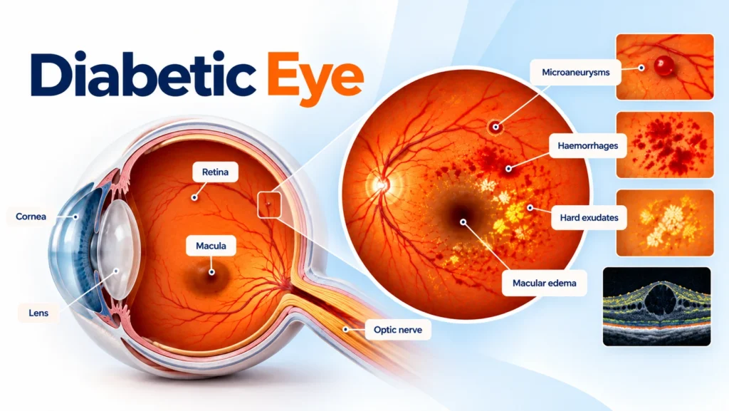
Which Tests Are Used in a Diabetic Eye Examination?
Not everyone needs every available retinal test. The ophthalmologist selects the appropriate investigations based on your medical history, symptoms and examination findings.
1. Dilated Eye Examination: Looking Directly at the Retina
A dilated eye examination is one of the main ways an ophthalmologist checks for diabetic retinopathy.
How does it work?
The doctor uses eye drops to widen your pupils, allowing a better view of the retina and its blood vessels. Specialised instruments are then used to examine the back of the eye.
The examination can reveal small areas of bleeding, damaged blood vessels, deposits associated with leakage and signs of abnormal new blood vessel growth.
Does it hurt?
The examination is generally painless. The drops may sting briefly, and your vision may remain blurry or sensitive to light for several hours.
A dilated examination provides a direct view of the retina and helps the doctor decide whether additional imaging is needed.
2. Fundus Photography: Capturing Images of the Retina
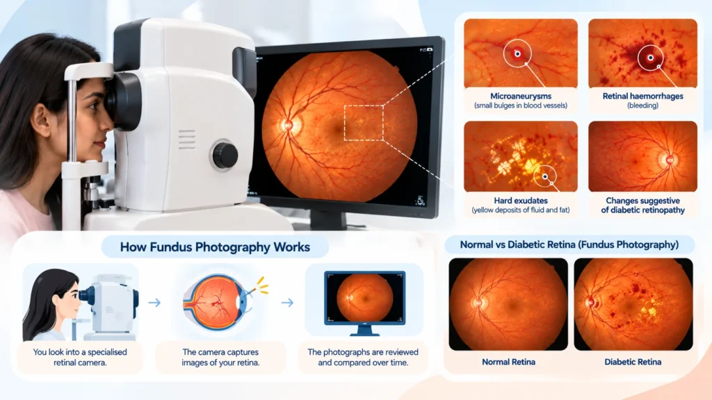
Fundus photography uses a specialised camera to capture photographs of the back of the eye. These images provide a visual record of the retina and can help identify changes associated with diabetic retinopathy.
How does it work?
You look into a retinal camera while it captures images of your retina. Depending on the equipment and the quality of the images required, dilating drops may be used.
The procedure is quick and non-invasive. The photographs can also be compared with earlier images to monitor changes over time.
What can it detect?
Fundus photography can help identify:
- Microaneurysms: Small bulges in retinal blood vessels that may be an early sign of diabetic retinopathy.
- Retinal haemorrhages: Small areas of bleeding caused by damaged blood vessels.
- Hard exudates: Yellowish deposits associated with leakage from retinal blood vessels.
- Other retinal changes: Findings that may suggest diabetic retinopathy or progression of existing disease.
Is fundus photography enough?
It can be an effective screening and monitoring tool, but it does not replace every part of a comprehensive eye examination. If images are unclear or abnormalities are detected, the doctor may recommend a dilated examination or additional imaging.
3. Optical Coherence Tomography (OCT): Checking for Retinal Swelling
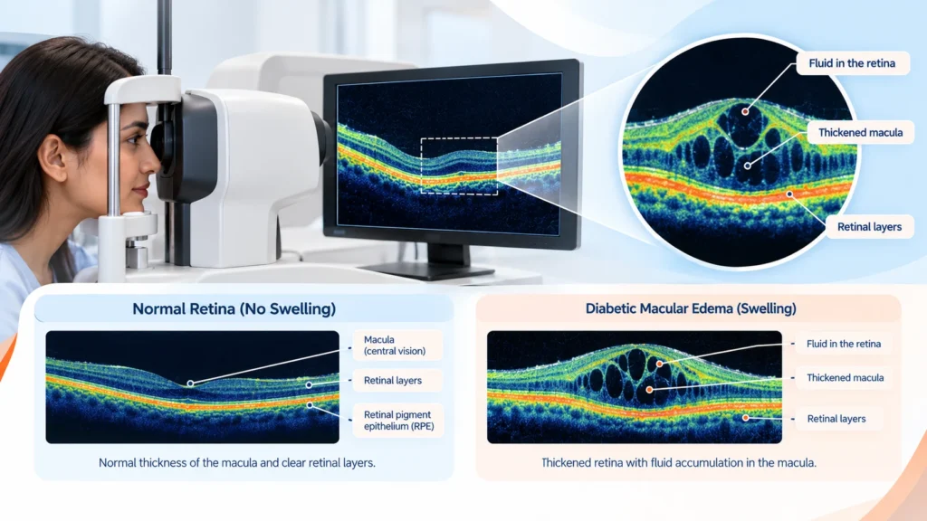
An optical coherence tomography scan, commonly called an OCT scan, creates detailed cross-sectional images of the retina. Unlike a conventional retinal photograph, it shows the different retinal layers and helps the ophthalmologist assess their thickness and structure.
OCT is particularly useful when diabetic macular edema is suspected.
How does it work?
You rest your chin on a support and look at a target inside the machine. The scanner uses light to capture detailed images without touching the eye.
The test is generally quick and painless. Dilating drops may or may not be needed, depending on the equipment and clinical situation.
What can an OCT scan detect?
It can help identify:
- Fluid within or beneath the retina.
- Thickening of the macula.
- Structural changes in the retinal layers.
- Features associated with diabetic macular edema.
- Changes in retinal structure over time or in response to treatment.
Why is OCT important?
A retinal photograph may show signs that raise concern about macular disease, but OCT provides a more detailed view of the retinal structure. This helps the ophthalmologist assess whether swelling is present and how extensive it is.
Not everyone with diabetes needs an OCT scan at every visit. It is recommended when the additional information is clinically useful.
4. Fluorescein Angiography: Examining Retinal Blood Flow
Sometimes, the ophthalmologist needs more information about how blood flows through the retinal vessels. Fluorescein angiography may be useful in these situations.
How does it work?
A fluorescent dye is injected into a vein, usually in the arm. A specialised camera captures photographs as the dye travels through the retinal blood vessels.
The images can reveal areas of leakage, reduced blood supply or abnormal new blood vessel growth.
When might it be recommended?
The doctor may consider this test to investigate blood vessel leakage, identify areas with reduced retinal blood supply, assess abnormal new vessels or obtain additional information for treatment planning.
Fluorescein angiography is not required for every diabetic eye examination. It is generally used when the results may help clarify the diagnosis or guide clinical decisions.
Temporary nausea or yellowish skin and urine discolouration can occur after the test. Serious allergic reactions are uncommon but possible, so tell your doctor about previous dye reactions and relevant medical history.
5. OCT Angiography (OCTA): Imaging Retinal Blood Vessels Without Dye
OCT angiography, or OCTA, is an advanced imaging technique that provides information about blood flow in small retinal vessels without requiring an injected dye.
How does it work?
The machine uses specialised OCT technology to produce images of the retinal microvasculature. The examination is non-invasive and generally painless.
What can it help identify?
OCTA may help the ophthalmologist assess changes in small retinal blood vessels, areas with reduced capillary blood flow and abnormal vascular patterns associated with diabetic retinopathy.
OCTA and fluorescein angiography provide different information. OCTA can visualise blood flow patterns but does not demonstrate leakage in the same way as fluorescein angiography. Image quality can also be affected by eye movement and other factors.
OCTA is a specialised investigation, not a routine requirement for every person with diabetes.
6. Visual Acuity and Eye Pressure Tests: Checking Overall Eye Health
A comprehensive eye examination may also include visual acuity testing and intraocular pressure measurement.
Visual acuity testing checks how clearly you can see letters or symbols at a specified distance. It helps assess your vision, but normal results do not rule out diabetic retinopathy.
Eye pressure measurement, also known as tonometry, measures the pressure inside the eye. It may form part of a broader assessment of eye health, including evaluation for glaucoma when appropriate.
These tests complement retinal examination and imaging, but neither provides a detailed assessment of the retina on its own.
Fundus Photography vs OCT: What Is the Difference?
Both tests help assess retinal health, but they serve different purposes.
| Feature | Fundus Photography | OCT Scan |
| What it produces | Photographs of the retinal surface | Cross-sectional images of retinal layers |
| Main purpose | Screening and documenting visible retinal changes | Assessing retinal structure and swelling |
| What it may detect | Microaneurysms, haemorrhages and hard exudates | Retinal thickening, fluid and structural changes |
| Common use | Screening and follow-up | Detailed assessment, particularly for suspected macular edema |
| Is it invasive? | No | No |
Which test is better?
Neither test is automatically better for every patient. Fundus photography shows visible retinal changes, while OCT provides more detailed information about the retinal layers. Depending on the findings, the ophthalmologist may recommend one or both tests alongside a dilated eye examination.
What Do Diabetic Retinal Examination Results Mean?
A retinal examination helps the ophthalmologist identify changes associated with diabetic eye disease. The findings can indicate whether the retina appears healthy, whether early changes are present or whether more advanced disease requires closer assessment.
Non-proliferative diabetic retinopathy
In this stage, retinal blood vessels may develop microaneurysms, haemorrhages and areas of leakage. The condition is classified as mild, moderate or severe according to the extent of these changes. Early disease may not affect vision noticeably.
Proliferative diabetic retinopathy
In this more advanced stage, abnormal new blood vessels grow in or around the retina in response to reduced blood supply. These fragile vessels can bleed and may contribute to serious complications, including retinal detachment.
Diabetic macular edema
Diabetic macular edema occurs when fluid accumulates in the macula. It can develop at different stages of diabetic retinopathy and may cause blurred or distorted central vision. An OCT scan is particularly useful for identifying retinal thickening and fluid associated with this condition.
For more information, read our guide to diabetic retinopathy.
How Often Should You Have a Diabetic Eye Examination?
The recommended screening interval depends on your diabetes type, previous examination results and individual risk factors.
- Type 1 diabetes: An initial comprehensive, dilated eye examination is generally recommended within five years of the onset of diabetes.
- Type 2 diabetes: An initial comprehensive eye examination is generally recommended at the time of diagnosis.
- After normal examinations: Screening every one to two years may be considered for some people with consistently normal retinal findings and blood glucose levels within their recommended targets.
- If diabetic retinopathy is present: Follow-up is generally needed at least annually, and more frequently if the disease is progressing or threatens vision.
- During pregnancy with pre-existing diabetes: Eye assessment before pregnancy or early in pregnancy is important, with further monitoring based on retinal findings and clinical advice.
These are general recommendations, not a personalised schedule. Your ophthalmologist may recommend more frequent examinations depending on your results and medical history. The American Diabetes Association’s 2026 Standards of Care provide further guidance on diabetic retinopathy screening.
When Should You Seek Urgent Eye Care?
Early diabetic retinopathy may not cause symptoms, which is why routine screening matters. However, certain changes in vision should not wait for a scheduled appointment.
Seek urgent eye care if you experience:
- Sudden loss or significant blurring of vision.
- A sudden increase in floaters.
- Flashes of light.
- A dark shadow or curtain across your vision.
New distortion of straight lines or persistent difficulty seeing clearly also warrants prompt assessment. These symptoms can have several causes, some of which require urgent treatment, and should not automatically be attributed to diabetes.
To support your eye health, work with your treating doctor to manage blood glucose, blood pressure and cholesterol, attend recommended retinal screenings, and follow your ophthalmologist’s advice. Good diabetes management can reduce the risk of diabetic retinopathy developing or progressing, but it cannot guarantee that eye complications will never occur.
Medical Disclaimer
This article is for general educational purposes only and does not replace professional medical advice, diagnosis or treatment. The appropriate tests and screening schedule should be determined by a qualified eye care professional based on your medical history and examination findings. Seek urgent medical assessment for sudden vision changes or vision loss.
References
- American Diabetes Association Professional Practice Committee for Diabetes. 12. Retinopathy, Neuropathy, and Foot Care: Standards of Care in Diabetes—2026. Diabetes Care. 2026;49(Supplement 1)–S276.
- Dubai Health Authority. Clinical Guidelines for Virtual Management of Type 2 Diabetes Mellitus.
- Emirates Diabetes Society. Consensus Guidelines for the Screening and Management of Diabetic Retinopathy in the Gulf Region.